Submitted by Christine Alexander on Thu, 17/03/2016 - 15:14
Centrosomes are the major microtubule organising centres (MTOCs) in somatic animal cells. They are localised at each pole of the mitotic spindle and persist throughout the life cycle of a cell. Each centrosome is composed of two barrel-shaped structures, known as centrioles, surrounded by electron dense material that contains proteins required for microtubule nucleation.
Towards the end of the nineteenth century, Theodor Boveri, who had provided one of the earliest descriptions of the centrosome, reported the presence of multiple spindle poles in tumour cells. Today we know that extra centrosomes are present in a wide range of tumours, both in early and advanced stages of disease, where they generally indicate a poor prognosis. However, the contribution of centrosomal amplification to oncogenesis remains unclear.
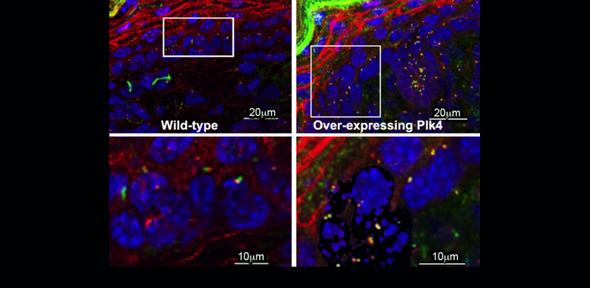
Members of the Glover Group have addressed this exact question, taking advantage of a transgenic mouse line that allows inducible over-expression of Plk4, the master regulator of centrosome formation. Elevating levels of Plk4 leads to formation of extra centrosomes, at the expense of primary cilia - small antenna-like structures templated by centrosome components that act to convey signals between cells. The net result is hyperplasia in several tissues, which is exacerbated by the additional loss of the tumour suppressor protein p53. p53 is non-functional in the majority of human tumours and mice lacking p53 are prone to tumour development. Interestingly, the Glover Group has observed that tumour appearance is accelerated when Plk4 levels are elevated in a p53 null background.
Now that the link between centrosome number and tumorigenesis has been established, the challenge will be to find the precise molecular connections between centrosomes the mitotic spindle they organise, and the p53 surveillance pathway.
The complete report of these findings is published in:
Coelho et al., (2015) Over-expression of Plk4 induces centrosome amplification, loss of primary cilia and associated tissue hyperplasia in the mouse. Open Biol. 5, 150209
Figure legend:
Cross-section of mouse backskin both wild-type and Plk4 over-expressing mice. DNA (in blue), centrioles and primary cilia (green) and Plk4 (in red) are shown. In the wild-type sections basal bodies and primary cilia are one per cell. However, after centriole over duplication the number of cells showing primary cilia in the epidermis is reduced.
