
Outreach Activities 2026
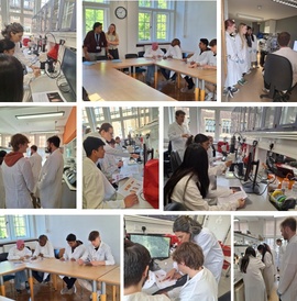
Magdalene College Easter Residential Sessions, 2026
We were delighted to host Year 12 students from schools in Liverpool and Wales in our labs for a hands-on science experience. For several of the students, this was their first visit to Cambridge, which made the experience all the more special.
During the sessions, students worked in a Drosophila research lab and gained practical experience in fly phenotyping, staining and imaging techniques, as well as Gram-positive and Gram-negative bacterial counterstaining. It was inspiring to see such enthusiasm and curiosity, and the sessions were a wonderful example of how hands-on research experiences can support and motivate the next generation of scientists.
The feedback we received included: “Students really loved the session in the Department and found it incredibly informative and insightful.”
More information on Magdalene College Easter Residentials 2026
Volunteers who supported the sessions: PhD student Maciej Zurowski, Dr James Earwaker Hammond, Dr Margherita Battistara, Dr Matt French, Dr Malaka De Silva, Dr Tom Pettini and Dr Juliette Nourry.
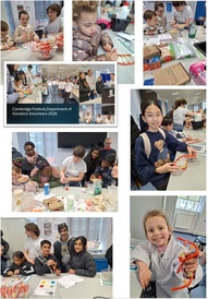
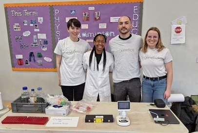
Cambridge Festival, March 2026
A big thank you to everyone who volunteered, helped out and visited the Dept of Genetics stand at the Cambridge Festival Family Day on Saturday 28 March. The dept had a fantastic presence, offering lots of interactive activities for children.
Feedback from the young participants included “I now know how DNA works", "It was fun and enjoyable especially liked the strawberry DNA it was interesting", and “so cool and fun definitely coming back again”.
Trumpington Meadows Primary SchoolSTEM event, March 2026
On 10 March, four members of the Department visited Trumpington Meadows Primary Schoolto support their STEM event.
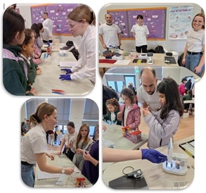
The activities prepared and delivered on the day included:
- DNA extraction from saliva – a hands-on experiment that allowed children to learn about DNA and visualise their own DNA in Eppendorf tubes.
- Handwashing activity – demonstrating the importance of good hygiene using “before and after” Petri dishes.
- Pipetting “Liquid Rainbow” – a fun and engaging pipetting activity introducing basic lab skills.
- Microscopy station – observing fixed slides through a portable microscope.
Volunteers:
Martin Santamarina Garcia, Mercy Suchanek, Juliette Nourry, and Robert Powles.